Lorem ipsum dolor sit amet, consectet eiusmod tempor incididunt ut labore e rem ipsum dolor sit amet. sum dolor sit amet, consectet eiusmod.
| Mon - Fri: | 8:00 am - 8:00 pm |
| Saturday: | 9:00 am - 6:00 pm |
| Sunday: | 9:00 am - 6:00 pm |


PET-CT (Positron Emission Tomography–Computed Tomography) is a cutting-edge imaging technology that combines metabolic and anatomical scans to provide highly accurate insights into various health conditions, especially cancer.
At Fusion PET-CT, we offer a range of PET-CT scans tailored to detect, stage, and monitor specific diseases with precision, aiding in better treatment planning and faster recovery.
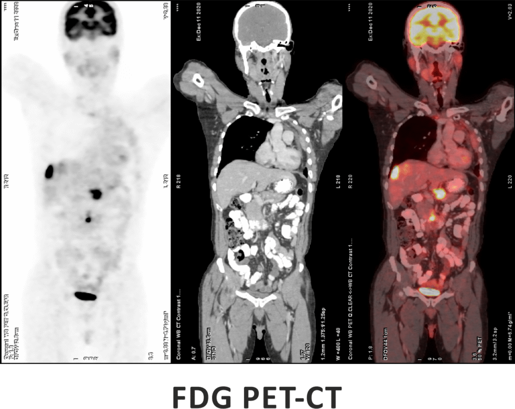
FDG PET-CT is one of the most advanced and widely used imaging techniques in modern medicine, especially for cancer evaluation. It works by detecting areas of increased glucose metabolism in the body, which is a common characteristic of active cancer cells, infections, and certain inflammatory conditions. By combining the metabolic insights from PET with the anatomical detail of CT, FDG PET-CT offers exceptional accuracy in locating and characterizing disease.
This scan plays a critical role in the diagnosis, staging, and treatment planning of a wide range of cancers. It also helps assess how well a tumor is responding to therapy and can detect recurrence at an early stage, often before it becomes visible on conventional scans.


68-Ga DOTANOC PET-CT is an advanced nuclear imaging technique specifically designed for the detection and evaluation of neuroendocrine tumors (NETs). It works by targeting somatostatin receptors, which are commonly overexpressed in these tumors. This scan provides high-resolution images that help in localizing the primary tumor, assessing the extent of disease spread, and evaluating suitability for treatments like Peptide Receptor Radionuclide Therapy (PRRT). It is particularly valuable in managing gastroenteropancreatic tumors, paragangliomas, pheochromocytomas, and other NET-related conditions.
68-Ga PSMA PET-CT is an advanced molecular imaging technique specifically designed for detecting prostate cancer. It targets the Prostate-Specific Membrane Antigen (PSMA), a protein that is highly expressed on prostate cancer cells. This scan offers exceptional sensitivity and accuracy, even at very low PSA levels, making it valuable for both initial staging and identifying recurrence. It also plays a critical role in guiding treatment decisions, especially for patients being considered for PSMA-targeted therapies.


Brain PET imaging is a highly advanced diagnostic tool used to assess the brain’s metabolic activity and function. It plays a crucial role in the early detection and evaluation of neurological conditions such as Alzheimer’s disease, various forms of dementia, epilepsy, and movement disorders like Parkinson’s disease. By analyzing how different areas of the brain process glucose, Brain PET helps distinguish between types of cognitive decline, assess disease progression, and guide treatment planning with greater accuracy than structural imaging alone.
Cardiac PET-CT is an advanced imaging technique that evaluates both the blood flow (perfusion) to the heart muscle and the viability of the myocardium (heart tissue). It is particularly useful in identifying areas of the heart that may still benefit from procedures like angioplasty or bypass surgery, even after previous heart damage.
Compared to conventional tests such as SPECT or treadmill stress tests, Cardiac PET-CT offers greater accuracy, faster imaging, and lower radiation exposure, making it ideal for high-risk or complex cardiac patients.

It combines two imaging methods to reveal both the structure of organs and how active those tissues are. This helps doctors detect disease early, understand how far it has progressed, and plan treatment with better accuracy.
It’s often recommended when doctors need more clarity about cancer, neurological issues, or certain heart conditions. It also plays an important role in checking how well ongoing treatment is working.
You’ll be with us for around two to three hours. This includes the tracer injection, a short waiting period, and the scan itself, which typically lasts 20 to 30 minutes.
The scan is painless. Apart from a small prick when the tracer is given, most people feel relaxed throughout. You just need to stay still during the imaging.
The tracer is used in very small, controlled amounts. It passes out of your body naturally within a few hours. Most patients do not experience any side effects.
Yes, a few simple guidelines help ensure accurate results. You may be asked to fast for a few hours, avoid heavy exercise, and follow special instructions if you have diabetes. All details will be shared when your scan is scheduled.
In most cases, you can. If anything needs to be paused or adjusted, our team will let you know in advance.
Please bring any previous reports, prescriptions, and your ID proof. If you’ve had earlier imaging studies, those help our specialist compare and report more precisely.
Reports are usually ready the same day or within 24 hours. If your doctor has requested an urgent review, we do our best to prioritise it.
Yes. You can go home immediately and continue with your routine. Drinking more water helps clear the tracer faster.
Many insurance plans cover PET-CT scans when medically indicated. Our team will guide you with paperwork, approvals, and cashless options depending on your insurer.
You can call us, submit an enquiry through the website, or visit the reception directly. We’ll help you choose the earliest available slot at the branch that’s most convenient for you.